Updated: September 4, 2010
Here's an experiment I want you to do. Close your eyes. Now, start gently rubbing your eyes with your hands, applying an ever greater pressure. Once you reach that bittersweet balance point between drowsy comfort and slight soft pain, maintain a steady pressure. Focus on the flashing images playing across your vision.
What do you see?

This is what I want to talk to you about: images created by physical pressure against your eyeballs rather than light photons. From what I've been able to gather, these images are almost universally similar, but my test pool has been relatively small, just a few friends and family members. Perhaps you can help unveil the mystery and set the truth free.
But first, let me tell you my version.
Fractal patterns
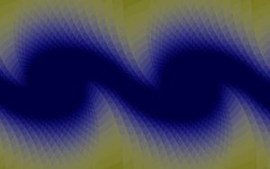
For me, the pressure against eyelids manifests in a static flicker of rectangles, alternating between yellow and purple, with an increasing frequency. I am fully aware that these would-be images are nothing more than my mind's interpretation of electric discharge in the retina caused by mechanical forces against the eyelids, but still, why only rectangles? And why yellow and purple?
The rectangles are sometimes spread across the entire vision and sometimes they form a kaleidoscope fractal-like pattern with several instances of rectangle areas. When this happens, in addition to the flicker, the rectangle areas also rotate slowly, probably induced by eye fluid motion.
Here's an approximate computerized interpretation of what I see. Not the most accurate, but something along those lines. This is hardly an attempt to show off my GIMP skills. Drawing this image was not a simple deal.

Physical answer
I don't think there's one. At least not a simple answer. I believe there's no mathematical equation that can translate eyeball movement into pictures, not without the use of a personal, intimate consciousness filter, anyway. But the fact we all see pretty much the same probably means something.
During my university studies, I did take a couple of biology courses, where we did focus quite a bit on how the human vision works. Technically, it comes down to photons hitting cone and rod cells and causing the 11-cis retinal to undergo photoisomeration to all-trans retinal, leading to signal transduction. Photons work best with eyes open, but even with eyelids shuttered, some of the particles do reach the retina, which explains the occasional flickers and dots caused by an odd discharge. Then, you can also cause similar effect by mechanical pressure.

But I don't see why the pressure should translate into a fractal-like pattern of rectangles. This is not a background vision noise, since it does not happen with eyes closed and no pressure applied. It might be a mapping of vision cells, saturated with clutter signal due to pressure, so what you're seeing is might be the topology of your own retina. And it could be nothing more than imagination.
If you have a theory, feel free to hypothesize.
Conclusion
No major conclusion, just a few big questions for you: What do you see? Are you like me, a rectangle person in yellow and purple? What kind of images does your mind fashion in response to mechanical pressure? Do you have a fancy idea that might explain the phenomenon?
If you've got a surreal eye moment to share, then feel free to send me an email and tell me about it. I'll post the most interesting or unique images (don't make things up!) as an update to this article.
Cheers!
Update, March 2011
Here's what Feefer had to say on this subject - unchanged, except just a tiny massage on some of the typos and such. Anyhow, a good explanation on what I've surmised above.
Interesting depiction of mechanical pressure (more on that, later). When I was a kid, I remember laying in bed in
total darkness, pressing on my eyes to see the kaleidoscope patterns (a bad idea, BTW; bad for the eyes). As a
graduate of UC Berkeley School of Optometry (known for its vision research), I can relate to what we were
taught.
FIrst off, you cannot actually see anything inside your eye (i.e. you cannot actually form images on the retina
from anything that's IN the eye), but only can experience the EFFECT that structures inside the eye have on
vision. Thus you cannot "see" the retina, or see the vitreous strands floating in your eye; you only can
perceive the effect these structures have on your vision. In the case of "floaters", we perceive them because
light from the outside world casts a SHADOW on the retina, so you're perceiving the shadowy imagery formed on the
retina
underneath.
There are certain circumstances where you can perceive the vascular structures lining the retina (vascular
arcades): I often tell patients to look for a branching pattern (which looks like tiny, delicate vessels on a
tree leaf) when I'm examining their eyes in a dark room, as I'm shining a bright light in their eye; many
patients will be able to "see" their own blood vessels, which represents a small minority of blood vessels inside
of the eye. Fortunately for us, the lion's share of retinal vasculature runs UNDERNEATH the retina, and not the
ones that can be perceived (which are on TOP of it); otherwise we wouldn't be able to see well, since the blood
vessels would block the light (as these small minority do).
Directed focal mechanical pressure on the eye can cause the retinal cells to misfire, since they are very
sensitive to ANY stimulation. Think about it: if the retina can detect the minuscule energy of PHOTONS hitting
the retina, it also can trigger as a result of the greater energy of mechanical pressure. Clinically, we
encounter this phenomenon when preforming scleral indentation, a procedure where we insert a dull metal probe
between the eyelid skin and orbital bone, applying gentle pressure indirectly on the globe to elevate the retina
layer underneath (e.g. when looking for a shallow retinal detachment). You can experience the perception at
home by closing your eyes and looking down as far as you can: gently place the eraser side of a pencil in the gap
on the outer surface or your upper eyelid, GENTLY pushing it into the space of the upper orbital bone and the
eyeball underneath the eyelid (NOT directly on the globe of your eye!). If you push, you should see a bluish
percept, corresponding to the area of the retina you're stimulating: look for it on the OPPOSITE side of the area
you're stimulating (you may remember that retinal imagery for the inferior visual field is handled by the
superior retina, i.e. it's inverted). In fact, sometimes people will "see" the point where the insertions of the
extra-ocular eye muscles are, as a result of flexing of the retina underneath those points.
In general, pressure elevation inside the eye (as caused by pressing your hands in your eyes) causes a decrease
in blood perfusion pressure to the retina, causing a transient (temporary) decrease in blood flow. The retina is
highly metabolic, with it's cells (cones and rods) having the highest rate of oxygen utilization (on a
weight-by-weight basis) of any cells in the body. Hence, the retina is very sensitive to a lack of blood flow,
and really needs oxygen to function properly; anything that causes a temporary disruption will cause the retina
cells to malfunction. And if the ischemia continues, those retinal cells will die within minutes, leading to
permanent vision loss if the blood flow isn't resumed.
This is partly why a fighter pilot pulling Gs notice that just before they pass out (faint), their vision will
black out, starting from the edges and moving inwards to the center of their vision. The blood flow to the
center of vision (the macula) is greatest, and that's why it's last to go. That higher rate of blood flow to the
macula also explains why the macula is damaged by conditions like macular degeneration, diabetic retinopathy,
etc.
I suspect the percept of kaleidoscope-like photopsias partly results from visual cortical activity, but it's a
bit like asking what the sound of one hand clapping is: it's impossible to tease out one, without considering
contribution of the other. For example, there are regions of the visual cortex in the brain which are responsible
for coding for geometric patterns (some quite complex), and I suspect the anoxia of the retina allows these cells
to "free-wheel" (run amok), in the absence of legitimate valid information coming from the retina.
I suspect individuals who have total cortical blindness won't see ANY kaleidoscope pattern (depending on the
depth of their affliction), while those who are blind in one eye from retinal disease (e.g. central retinal
artery occlusion, leading to almost total loss of vision in one eye) will notice these types of patterns, and
even find them quite distracting, as that kaleidoscope pattern is projected on the vision formed by their
remaining "good" eye, thus interfering with its vision.
Hope this helps.